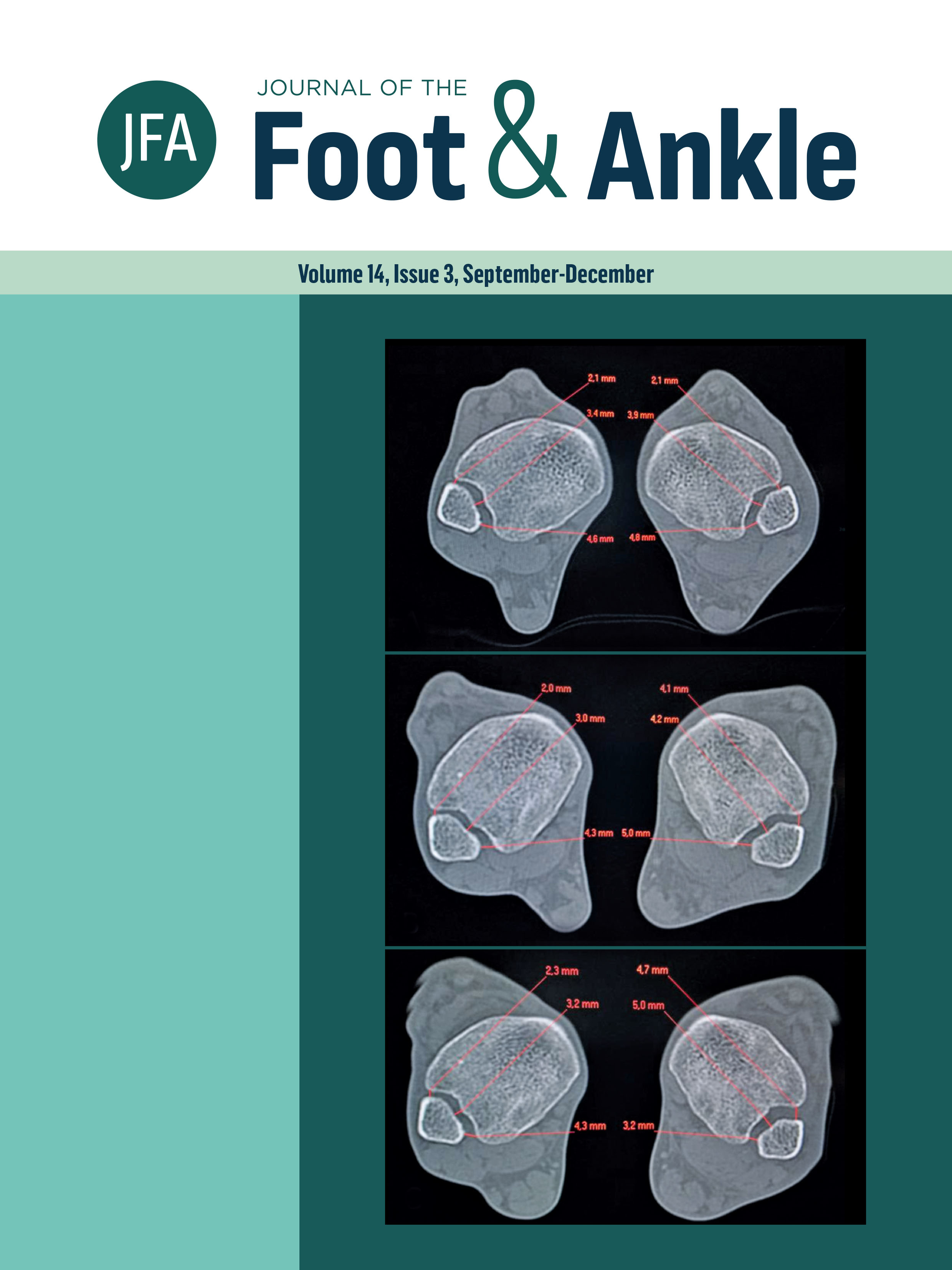
The interobserver reliability of first metatarsal rotational component of axial sesamoid radiographs in hallux valgus
DOI:
https://doi.org/10.30795/jfootankle.2020.v14.1196Keywords:
Bunion, Metatarsal bones, Foot deformities, Pronation, RotationAbstract
Objective: Hallux valgus is a progressive triplanar deformity of the forefoot with an important rotational component (RC) in the first metatarsal, which has been associated with recurrence. There is controversy about using weight-bearing vs. non-weight-bearing radiographs in RC measurement. This study aims to assess interobserver reliability for RC of the first metatarsal using a non-weight-bearing sesamoid view, as well as to correlate the hallux valgus angle, intermetatarsal angle, distal metatarsal articular angle (DMAA) and sesamoid position regarding RC. Methods: An observational, cross-sectional and descriptive study was conducted with 81 feet from 48 patients (66.6% female). RC was evaluated regarding the first metatarsal proximal shaft in non-weight-bearing axial metatarsal radiographs and weight-bearing anteroposterior radiographs. Measurements were taken independently by two foot and ankle subspecialists and an orthopedic resident, all of whom were blinded. Results: Statistically significant intraclass correlations (p = 0.02) were obtained for first metatarsal RC assessment among the three observers (95%CI 0.01–0.65; Cronbach’s α =0.41) in non-weight-bearing axial metatarsal views. Significant correlations (Spearman ρ) were also found for hallux valgus angle (p = 0.04) and DMAA (p = 0.01), and non-significant correlations were found for intermetatarsal angle and sesamoid position (p > 0.05). Conclusion: The significant correlations between hallux valgus angle and DMAA for RC suggest that RC is isolated from the first metatarsal bone structure. This practical assessment method may isolate the first metatarsal head RC regarding the proximal metatarsal in the metaphyseal region and could be useful in centers where weight-bearing CT scans are not available. Level of Evidence IV; Therapeutic Studies; Case Series.


