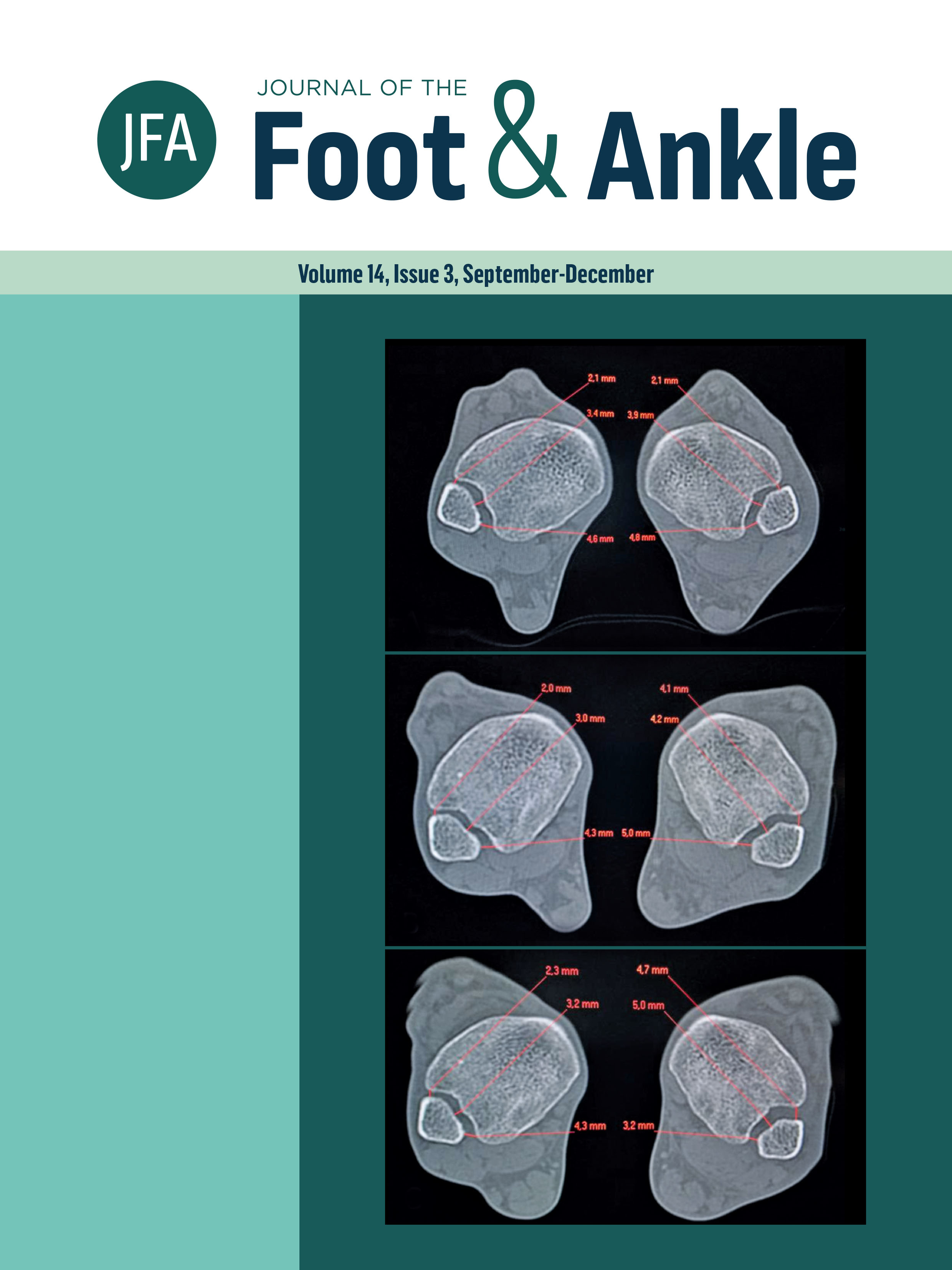
Hindfoot alignment using weight-bearing computed tomography
a new measurement for pes cavovarus
DOI:
https://doi.org/10.30795/jfootankle.2020.v14.1214Keywords:
Talipes Cavus/diagnostic imaging, Weight-Bearing/physiology, Tomography, X-Ray computed/methods, Bone malalignmentAbstract
Measurement of hindfoot malalignment and flexibility is essential for treatment decision-making in cavovarus foot deformity. Weight-bearing computed tomography (WBCT) shows greater diagnostic accuracy and allows the study of osteoarticular alignment in the physiological upright position. The most commonly used method for measurements on WBCT scans is the foot and ankle offset (FAO), which is based on the structural tripod of the foot: the calcaneus and the first and fifth metatarsal heads. During the Coleman block test, the first metatarsal head is not resting on the ground and, therefore, does not represent the physiological support of the tripod. We describe a new measurement, the forefoot/hindfoot offset (FHO), for assessing hindfoot alignment on WBCT scans. Level of Evidence V; Diagnostic Studies; Expert Opinion.