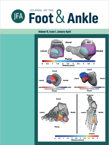
Weight-bearing CT Hounsfield unit algorithm assessment of calcaneal osteotomy healing. A prospective study comparing metallic and bio-integrative screws
DOI:
https://doi.org/10.30795/jfootankle.2022.v16.1630Keywords:
Absorbable implants, Biocompatible materials, Calcaneus, Fracture healing, Orthopedic fixation devices, OsteotomyAbstract
Objective: To investigate the capability of biointegrative screws to achieve similar radiographical healing outcomes to metallic screws, measured using Hounsfield Unit (HU) algorithms, in medial displacement calcaneus osteotomies (MDCO). Our main hypothesis is that both implant methods would demonstrate comparable results. Methods: In this prospective comparative study, patients undergoing MDCO were allocated to either a biointegrative or a metallic group. Surgeon, primary diagnosis, technique, and displacement were the same for both groups. Patients were assessed using weight-bearing computed tomography preoperatively and at weeks 2, 6, and 12 postoperatively. A 40x40x40 mm cube was centered on the osteotomy site, defining a volume of interest (VOI). Image intensity (Hounsfield Units) profiles along lines perpendicular to the osteotomy line and crossing it were recorded. Graphical plots of the HU distributions were generated for each line and then used to calculate the HU contrast. Results: Three patients were allocated to the metallic group (age: 50.66; BMI: 27.78) and three to the biointegrative group (age: 47.33; BMI: 39.35). At two weeks, mean HU intensity was lower in the metallic group on the center (403.25 vs. 416.28; p=0.312) and superior lines (438.97 vs. 497.92), but not on the inferior line (513.24 vs. 386.57; p<0.001). At six weeks, the mean HU intensity was higher in the biointegrative group on the center line (p<0.001) and the superior line (p=0.018). At 12 weeks, the metallic group presented lower HU values on the center (p<0.001) and inferior (p<0.001) lines, but higher values on the superior line (p=0.010). Contrast was higher in the metallic group patients in the second (p=0.034) and 12th weeks (p=0.049). Conclusion: Bone healing radiographical status results were similar for metallic and bio-integrative screws. Maximum HU values were equivalent, indicating comparable results at the osteotomy sites. However, the biointegrative group had lower contrast, portraying lower variability of bone density over the area. Level of Evidence II; Prospective Comparative Study.
Downloads
Published
How to Cite
Issue
Section
License
Copyright (c) 2022 Journal of the Foot & Ankle

This work is licensed under a Creative Commons Attribution-NonCommercial 4.0 International License.


